There’s a particular kind of frustration you only get in neuroscience when you know something is happening, you can feel the pattern in the data, and yet your instruments refuse to show it cleanly. The brain is electric, yes—but it is also magnetic in a way most people never internalize. Every action potential is not just a voltage spike; it’s a moving current, and moving currents whisper magnetic fields into the space around them. These fields fall below intuitive physical scale: femtotesla to picotesla depending on geometry, distance, synchrony, and the structure you’re trying to observe. But “small” doesn’t mean “meaningless.” It means your sensor has to be honest, quiet, and unbelievably sensitive.
For decades, neuroimaging has been a trade between compromises. EEG is fast, but spatially blunt—sharp in time, fuzzy in place. fMRI is spatially elegant, but temporally slow—high-resolution spatial reconstructions that inherently lag underlying neural events. MEG sits in an awkward middle: very compelling physics, very expensive hardware, and historically tied to cryogenics and rooms that feel like they were built for an experiment, not a hospital. In labs, you can tolerate that. In the real world, you start noticing how quickly “non-invasive brain mapping” becomes “non-invasive under ideal conditions.”
My interest in this area did not come from promotional narratives about “quantum advantage,” but from observing measurement groups treating diamond defects as practical instrumentation. The shift was seeing serious groups treat diamond defects as measurement devices—quietly, almost stubbornly—while everyone else was still describing quantum sensors as a category without committing to what makes them special. Nitrogen-vacancy (NV) centers in diamond are not a metaphor. They are a specific physical defect—one nitrogen atom next to a missing carbon site in the diamond lattice—that behaves like a controllable quantum system you can initialize, manipulate, and read out optically. That last word matters: optically. Not cryogenic wiring. Not heavy coils wrapped around fragile electronics. Light in, light out, with microwave control in between. [1]
When you take that seriously, NV center brain mapping stops sounding like a headline and starts sounding like instrumentation: a room-temperature quantum magnetometer that can sit close to tissue, measure vector fields with micrometer-scale locality, and—under the right conditions—track neural dynamics without touching the neurons with electrodes. Some of what’s been demonstrated is still ex vivo or in controlled environments. Some of it is closer to pilot-stage than clinical reality. But the underlying physics is real, and the engineering trend is not moving backward.
The discussion that follows examines NV sensing from first-principles physics and connects those mechanisms directly to neuroscientific measurement requirements. The emphasis remains on linking lattice-scale behavior with biological signal environments, since practical insight emerges at the interface between controlled quantum models and the variability inherent in living systems.
The Physics of NV Centers: Why Diamond Defects Became Quantum Sensors
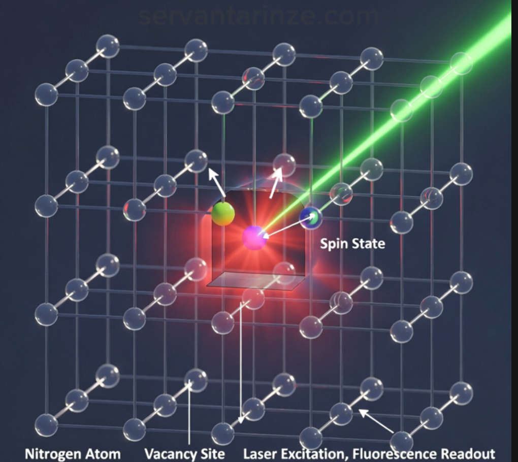
Describing NV centers as simple magnetic probes obscures the physical mechanisms that make them viable sensing platforms. The reason they became a magnetometry platform is that the defect hosts an electron-spin system with a structure that you can drive and read out in a way that survives room temperature. The commonly used charge state is NV−, and in that state the ground electronic configuration forms a spin triplet: S = 1. In practical terms, there are three spin sublevels—often labeled by the magnetic quantum number ms = 0, ±1—that behave differently under magnetic fields. [2]
Even before you apply any external magnetic field, the NV’s spin states are not perfectly degenerate. There’s a built-in splitting (the “zero-field splitting”) between the ms = 0 level and the ms = ±1 levels. When an external magnetic field is present along the NV axis, the Zeeman interaction shifts the energies of the spin sublevels in a field-dependent way. That shifting is not an abstract detail—it is the sensing mechanism. Your magnetic field literally changes the resonance condition of the defect.
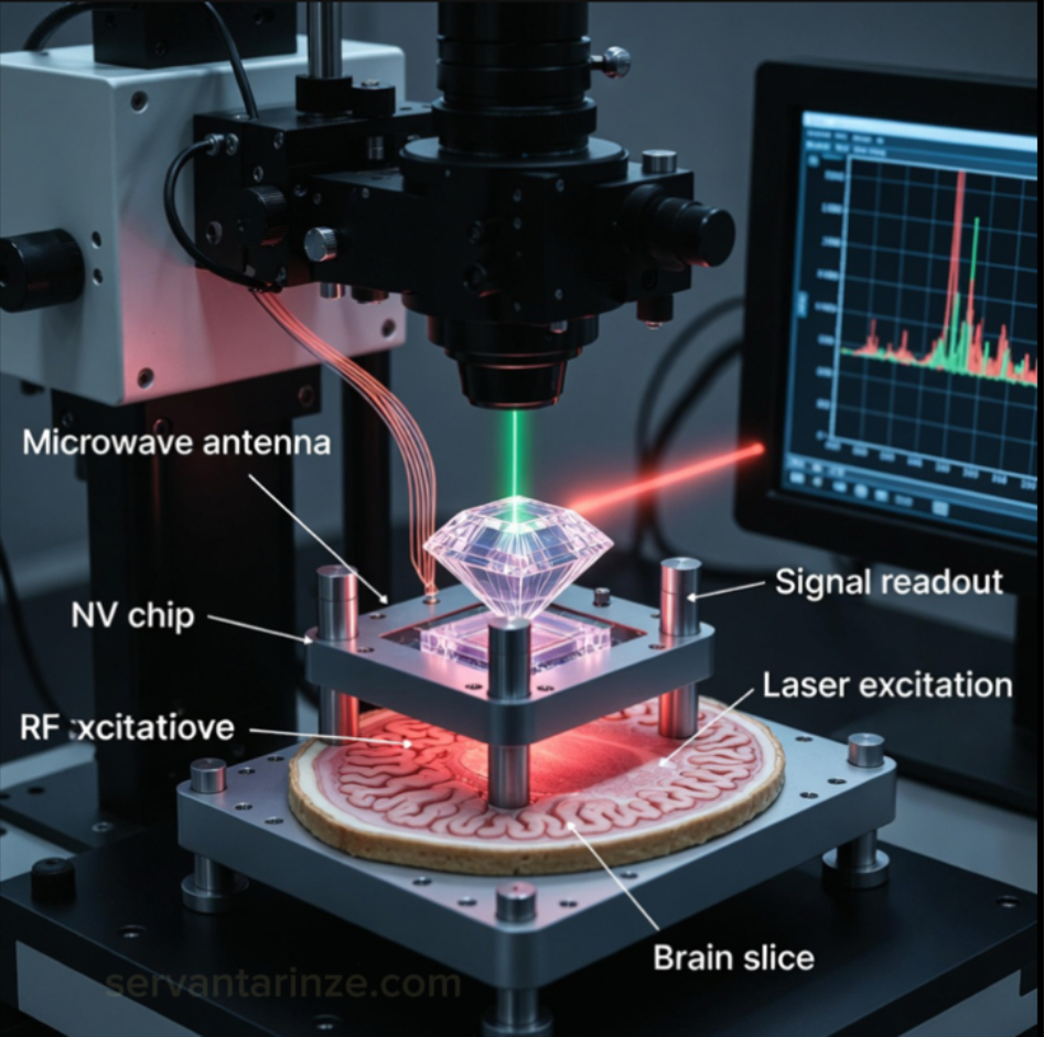
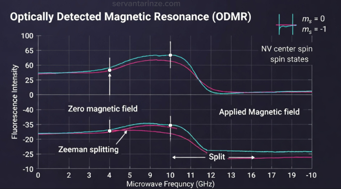
A defining advantage of the platform emerges in the optical initialization and readout pathway: you can initialize and read out this spin state with light. Under green laser excitation, the NV emits red fluorescence, and the fluorescence intensity depends on which spin state you’re in. That spin-dependent fluorescence contrast is what turns “a quantum state” into “a sensor output.” If you drive the system with microwaves and sweep the frequency, you can observe dips in fluorescence when the microwave matches a transition between spin sublevels. That’s optically detected magnetic resonance—ODMR—and it is the workhorse of NV magnetometry. [3]
What you measure in ODMR is not merely a spectrum. You’re measuring a spectrum that moves when the magnetic environment changes. A neuron firing nearby changes currents; currents generate magnetic fields; fields shift resonance; resonance shifts fluorescence. At a distance, those fields are tiny, but the NV does not care about your intuition. It cares about resonance. Resonance behavior therefore provides a precise accounting of magnetic perturbation.
There’s a second reason diamond keeps showing up in serious sensor conversations: coherence. The NV spin can maintain quantum coherence for surprisingly long times even at room temperature—especially in high-purity diamond and with careful control sequences. Those coherence times (often discussed through T2 and related measures) set the ceiling for sensitivity. A longer coherence time gives you a narrower effective linewidth in sensing, which improves your ability to detect small shifts caused by weak magnetic fields. Improved material purity and control protocols directly enhance signal stability and detectability—and suddenly “tiny neural fields” stops being a joke. [4]
But the physics doesn’t give free miracles. NV sensing always lives inside trade-offs. A single NV center can offer nanometer-scale proximity sensing, but your signal is photon-limited and painfully local. An ensemble of NV centers—many defects addressed at once—gives you far higher sensitivity and the possibility of wide-field imaging, but you trade away some locality and introduce averaging effects. This single-center versus ensemble trade-off remains a persistent structural consideration in experimental design, and it maps almost perfectly onto neuroscience itself: do you want single-neuron truth, or population-level stability? Most of the time, you want both, and you settle for a ladder of approximations.
Conceptual comparisons to macroscopic magnetic instruments should be treated cautiously. NV centers do not respond to large uniform fields in the manner of classical compasses; instead, they transduce extremely weak local perturbations into optical contrast variations. Their operational role is therefore better understood as localized magnetic transduction rather than directional indication.
And if the sensor is a microphone, neuroscience is a crowded room. The magnetic field you want is not the only field present. There are background fields, environmental noise, mechanical vibration, temperature drift, and the messy electromagnetic reality of living systems. The reason NV sensing is still taken seriously in this context is that ODMR is not just a detection trick—it is a tunable measurement protocol. You can engineer pulse sequences, choose measurement axes, and move between DC and AC magnetometry regimes depending on what kind of neural signal you’re hunting. That flexibility, in practice, is the bridge between physics elegance and biological usefulness.
From Spin Physics to Measurement: How NV Magnetometry Actually Works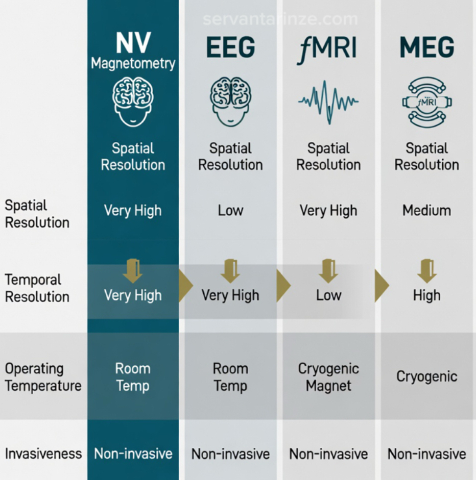
The transition from “interesting defect physics” to “usable sensor” happens when resonance shifts become measurable quantities. In NV magnetometry, what you ultimately observe is a change in fluorescence intensity as microwave frequency is tuned across the spin transition. The position of that resonance peak—or dip, depending on how you plot it—is directly related to the magnetic field projected along the NV axis. You’re not measuring the field in a classical probe sense. You’re inferring it from quantum energy level changes that light translates into contrast.
That distinction matters because it explains both the power and the fragility of the technique. The sensitivity you achieve depends on optical collection efficiency, photon shot noise, spin contrast, and coherence time. These parameters correspond directly to engineering limitations governing practical sensitivity. A microscope objective with poor collection efficiency is not a minor inconvenience; it directly worsens magnetic sensitivity. A diamond sample with residual paramagnetic impurities shortens coherence and raises your noise floor. The device is not separate from the measurement. It is the measurement.
In simplified form, the magnetic sensitivity relationship that experimentalists often lean on can be expressed conceptually as something proportional to:
Sensitivity ∝ 1 / √(contrast × photon rate × coherence time)
No one runs a lab from that single expression, yet it captures the governing physical relationship: more photons, better spin contrast, longer coherence — you hear quieter magnetic signals. This is why ensemble NV systems push optical throughput so aggressively, and why nanofabricated diamond structures are designed to funnel emitted photons toward detectors rather than letting them scatter uselessly into the lattice.
Once microwave control enters the loop, the NV center becomes dynamically tunable. Continuous-wave ODMR gives straightforward spectral shifts. Pulsed sequences allow selective filtering of temporal frequencies in the magnetic signal. Echo-based protocols suppress environmental noise. Vector magnetometry configurations exploit differently oriented NV axes in the crystal to reconstruct full field directionality rather than just magnitude. These adjustments represent structural modifications to measurement capability; they transform the defect from a static probe into something programmable, something you shape to the experiment.
This programmability is where comparisons to classical sensor technologies become meaningful. NV magnetometry is often introduced by contrasting it with SQUID systems or optically pumped magnetometers, but the comparison only becomes useful when grounded in what each technology prioritizes.
| Technology | Operating Conditions | Strength | Limitation | Relevance to Brain Mapping |
|---|---|---|---|---|
| NV Centers (Diamond) | Room temperature, optical readout | Nanoscale proximity, tunable protocols | Signal strength trade-offs, depth limits | Localized neural field sensing, scalable imaging |
| SQUID | Cryogenic superconducting | Extreme sensitivity | Bulky, expensive infrastructure | Established MEG standard |
| OPM | Heated vapor cell | Portable MEG advances | Spatial resolution constraints | Wearable neuroimaging direction |
| EEG | Room temperature electrodes | Temporal precision | Spatial ambiguity | Widely accessible monitoring |
| fMRI | Cryogenic superconducting magnet | Spatial structure mapping | Indirect metabolic signal | Functional localization |
I hesitate to frame this as a competition, because the neuroscience ecosystem rarely replaces tools outright. It layers them. NV sensing enters that ecosystem differently. Its advantage isn’t about dethroning SQUID sensitivity or EEG accessibility; it’s about proximity and modality. Magnetic fields decay quickly with distance. A sensor that can sit millimeters—or micrometers—from neural current sources is listening to a fundamentally different signal landscape than one meters away inside a cryogenic helmet.
That closeness is what makes discussions of NV center brain mapping credible rather than aspirational. Diamond probes integrated into microscopy platforms have already recorded signals correlated with neuronal activity in tissue preparations. Ensemble surfaces patterned for wide-field readout are pushing toward spatial mapping across slices. Parallel developments in ambient-condition magnetometry hint at scenarios where bulky shielding is less dominant than it once was. These are incremental shifts, not revolutions, but revolutions are built from enough increments accumulating in the same direction.
Still, physics imposes boundaries. The magnetic field from neural ionic currents weakens rapidly with distance, and biological environments are noisy. NV sensors do not magically erase Maxwell’s equations. The strength of the platform lies instead in its flexibility — its ability to approach the source, adapt measurement protocol, and operate without cryogenic overhead. That combination is precisely what neuroscience instrumentation has historically struggled to unify.
Which leads naturally to the next question — and the more biological one: how those neural currents produce measurable magnetic structures at all, and how NV platforms translate action potentials into maps rather than isolated signals. That transition from isolated measurement to spatial reconstruction is where the sensing physics meets neural geometry.
Magnetic Whispers of Neurons: Translating Biology into Measurable Fields
Clarifying the signal origin requires examining the electromagnetic consequences of neuronal current flow. Neural activity is electrical, not magnetic, but the two are inseparable. Whenever ions move through an axon during an action potential, they generate a current, and that current produces a magnetic field encircling the conductor according to Maxwell’s equations. These fields are extraordinarily small — typically tens of femtotesla to picotesla depending on geometry and distance — but they carry structural information about timing, direction, and synchronization that electrical recordings alone do not fully preserve.
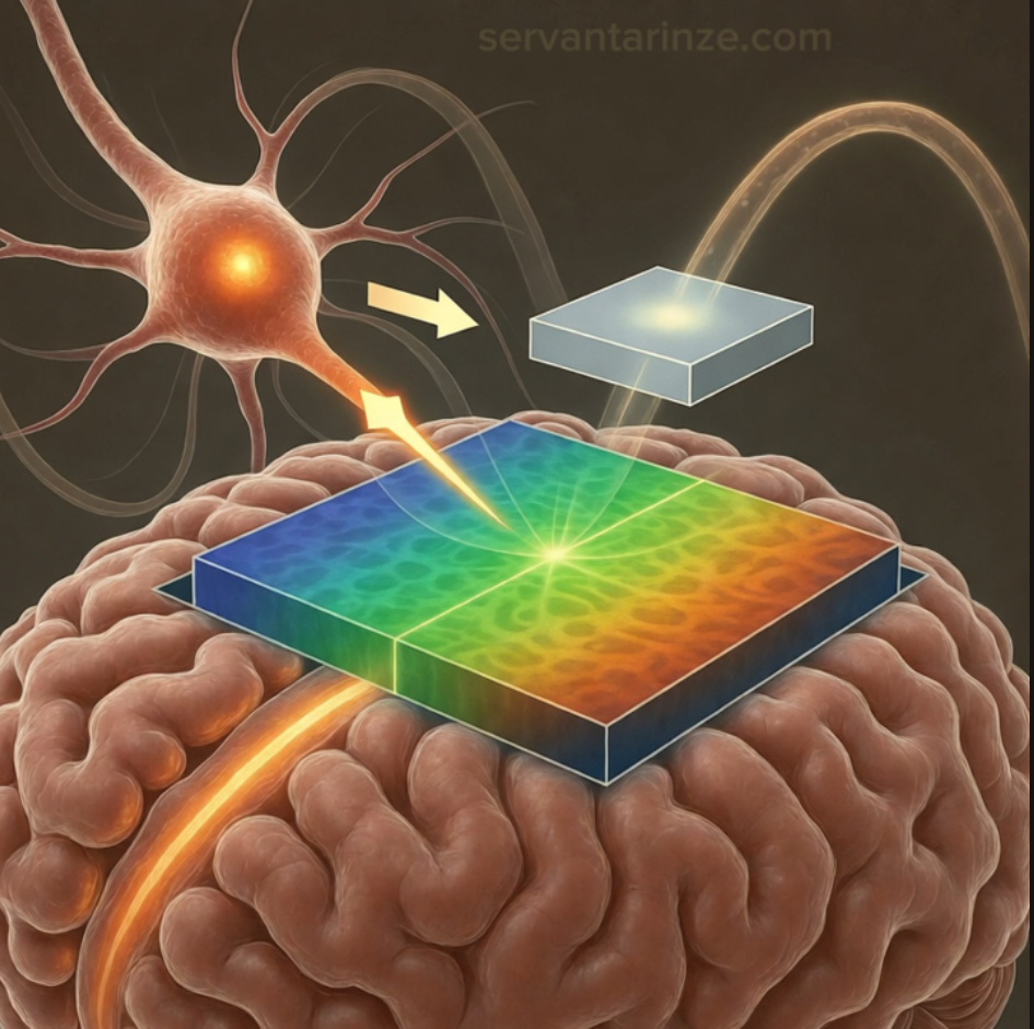
Traditional magnetoencephalography systems measure these fields from centimeters away, forcing them to interpret a heavily attenuated and spatially blended signal. NV-based approaches pursue the opposite philosophy: proximity. By positioning diamond sensors within micrometers of neural tissue or integrating them into microscopy stages, the measurement captures localized field structure before it disperses into collective background noise. This difference in distance transforms not just signal strength but interpretability. It changes the measurement from reconstruction-heavy inference into something closer to direct observation of current geometry.
In experimental settings, this proximity allows magnetic signatures from axonal propagation or synchronized network bursts to be distinguished without the large-scale averaging imposed by distant detectors. Cultured neuronal networks and brain slices have already demonstrated field patterns correlated with stimulation events, revealing spatial propagation dynamics that align with electrophysiological recordings yet provide independent validation through magnetic observation. Dual-modality correlation between electrical and magnetic measurements provides strong validation of signal attribution — two measurement paradigms converging on the same biological phenomenon from entirely different physical mechanisms.
Sensing Configurations: From Single Defects to Collective Ensembles
The architecture of NV sensing determines what scale of neural behavior becomes accessible. A single NV center offers nanometer spatial precision and can probe extremely localized magnetic environments. This regime is valuable when investigating subcellular structures or interactions with magnetic nanoparticles, but its photon collection rate limits raw sensitivity. In contrast, ensemble NV layers sacrifice spatial pinpointing in exchange for signal strength by averaging across millions of defects. The ensemble behaves less like a microscope tip and more like a distributed imaging surface capable of capturing broader activity patterns.
These configurations function as complementary measurement regimes occupying different resolution tiers. Single-defect measurements lean toward fundamental biophysics and nanoscale interaction studies. Ensemble platforms align with tissue-level imaging where sensitivity and field coverage dominate. Increasingly, hybrid architectures — patterned nanopillars, wide-field arrays, or structured illumination geometries — blur this distinction, attempting to extract sensitivity without surrendering localization.
Three principal detection modes appear repeatedly in neuroscience-oriented experiments. Frequency-shift ODMR remains the most intuitive, identifying resonance displacement caused by local magnetic fields. Relaxometry approaches monitor spin relaxation time changes induced by fluctuating environments, capturing dynamic processes that static resonance measurements may miss. Vector magnetometry combines multiple NV orientations to reconstruct directional field components, enabling geometric mapping rather than scalar intensity readings. Each mode observes neural activity differently, and selecting between them is less about preference than experimental intent.
The spatial-temporal envelope achieved by these techniques sits in an intriguing region between classical neuroimaging extremes. Spatial resolution can approach micrometer scales under microscopy integration, while temporal responsiveness can track microsecond-scale variations in magnetic fluctuations. This places NV sensing between electrode-based electrical recordings and large-scale imaging systems, bridging observational gaps rather than replacing existing modalities. It is this bridging role that gives the technology relevance beyond novelty.
Mathematically, discussions of sensitivity often return to scaling relations connecting contrast, photon statistics, and coherence duration. While detailed modeling varies across implementations, the essential intuition persists: improved photon collection and longer spin coherence extend the detectability of weak neural fields. Advances in diamond engineering and optical design are therefore not peripheral technicalities; they directly expand the biological phenomena accessible to measurement.
From a research perspective, what stands out is not just detection capability but interpretive fidelity. Neural systems exhibit complex spatial propagation patterns, and sensors that resolve vector structure rather than scalar magnitude preserve directional information crucial for understanding connectivity and signaling pathways. NV platforms, when configured appropriately, capture this directional nuance in ways traditional macroscopic sensors cannot.
These capabilities form the conceptual bridge into the next stage of the discussion — the empirical demonstrations and engineering milestones that have emerged across recent years. Physics principles alone do not establish credibility; what matters is how those principles manifest in laboratories measuring real tissue, real signals, and real noise environments. That is where NV sensing shifts from theoretical promise into documented experimentation.
Laboratories, Not Speculation: Where NV Brain Sensing Actually Stands
Discussion of quantum sensing becomes hollow unless grounded in measured outcomes, and over the past few years the NV–neuroscience intersection has quietly accumulated experimental weight. Published experimental literature reflects gradual improvement in signal fidelity rather than abrupt performance discontinuity and instrument stability. The emphasis has shifted from proving detectability to improving practicality — lowering environmental constraints, scaling sensor areas, and extracting signals that were previously buried in background variation.
One milestone frequently cited in magnetometry circles emerged from work exploring ambient-condition magnetoencephalography prototypes. Efforts from research groups including Tokyo Institute of Technology demonstrated sensitivity on the order of single-digit picotesla per root hertz without cryogenic shielding. That detail matters more than the number itself. Removing cryogenics and heavy shielding removes architectural friction that historically kept magnetometry confined to specialized facilities. It signals an engineering trajectory toward portability rather than institutional immobility.
Parallel advances have unfolded in microscopy-scale experiments where NV layers positioned beneath biological samples recorded magnetic signatures associated with axonal signaling. Brain slice preparations — particularly those examining corpus callosum pathways — showed measurable field correlations with induced activity, confirming theoretical predictions about current-induced magnetic emission. These measurements did not claim full in vivo mapping, and responsible interpretation avoids overstating them, yet they demonstrated something foundational: neuronal magnetic signatures are experimentally accessible through diamond-based quantum sensors operating at ambient temperature.
Wide-field imaging architectures strengthened this direction by trading single-point precision for spatial coverage. Instead of probing one coordinate at a time, planar NV ensembles captured distributed magnetic textures across tissue surfaces. Patterned nanopillar geometries increased fluorescence collection efficiency while maintaining local field sensitivity, producing sharper magnetic contrast maps. When I first encountered these images, their significance lies in structural correspondence rather than visual novelty — propagation shapes resembling electrophysiological wavefronts rendered in an entirely different modality.
Examples That Shifted the Conversation
Experimental recordings from cultured neuronal networks have provided additional confirmation. Controlled stimulation within engineered neural cultures generated synchronized magnetic responses observable through ensemble NV platforms. These observations paralleled electrode recordings while revealing spatial gradients invisible to electrical probes. The implication was subtle but important: magnetic observation supplements rather than duplicates electrical measurement, capturing complementary information about current distribution geometry.
Investigations involving magnetically active nanoparticles in neural environments have also leveraged NV sensing for biological characterization. Studies examining magnetite distributions within brain tissue demonstrated detection capability sensitive enough to resolve particle-induced field variations. While these experiments sit slightly adjacent to direct neuronal mapping, they reinforce the broader point that NV magnetometry functions robustly within biological complexity rather than only under idealized conditions.
Animal-model explorations have cautiously approached shallow in vivo contexts, where sensors positioned near cortical regions monitored induced activity signatures. These attempts remain early-stage and should be described as exploratory rather than transformative, yet they show directional progress toward bridging laboratory preparation and living-system observation. Each incremental improvement in sensor stability or optical efficiency shortens the distance between controlled experiment and clinical relevance.
Reviews emerging through interdisciplinary literature across physics and biomedical journals increasingly frame NV magnetometry not as speculative instrumentation but as an evolving member of the neuroimaging ecosystem. The narrative tone has changed. Instead of asking whether neural fields can be detected, authors debate optimization strategies, calibration reliability, and integration pathways with optical microscopy and computational filtering. That transition from feasibility discussion to implementation refinement is often where technologies quietly mature.
Visualization and Interpretation of Magnetic Neural Landscapes
Magnetic field mapping outputs carry interpretive weight only when contextualized alongside established imaging references. Comparative overlays aligning NV-detected propagation patterns with known anatomical structures help validate spatial correspondence. ODMR spectra showing field-dependent resonance shifts illustrate the measurement mechanism directly, reinforcing confidence that recorded variation arises from physical interaction rather than optical artifact.
When examining these visual outputs I find it useful to compare them mentally with classical imaging limitations. EEG compresses spatial detail; fMRI dilutes temporal immediacy; cryogenic MEG imposes infrastructural distance. NV visualizations inhabit a different observational niche — closer proximity, moderate field coverage, and compatibility with laboratory optical instrumentation. They do not solve every imaging problem, but they widen observational bandwidth across scales that were previously disconnected.
Perhaps the most meaningful conclusion drawn from these collective demonstrations is methodological rather than technological. Quantum sensing has entered biological experimentation not through abrupt disruption but through gradual assimilation. Each laboratory result contributes a fragment of credibility, and taken together they form an empirical foundation strong enough to justify continued investment and interdisciplinary attention. This accumulation of evidence is what shifts NV sensing from theoretical enthusiasm into measured scientific presence.
Where This Matters: Neuroscience Applications and Expanding Interfaces
Once the physics proves reliable and instrumentation stabilizes, the question shifts from capability to relevance. What becomes clear when following ongoing collaborations between sensing physicists and neurobiologists is that NV magnetometry is rarely framed as a replacement for established modalities. It enters the landscape as a complementary observer — offering access to spatial and temporal regimes that neither electrophysiology nor macroscopic imaging comfortably occupy. Its value emerges not from dominance but from filling observational gaps that persist despite decades of technological refinement.
One of the most compelling directions involves mapping neural dynamics within controlled biological models. Tissue cultures and organoid systems already serve as experimental stand-ins for studying connectivity development and pathological evolution. Magnetic sensing adds a layer of insight into current propagation patterns that electrodes may spatially undersample. Because NV detection does not require physical penetration of the sample, repeated observation becomes less invasive, preserving fragile biological conditions over extended monitoring cycles.
Disease-model experimentation illustrates another meaningful application space. Neural degradation associated with conditions such as epileptiform activity or plaque-related disruption introduces measurable shifts in current pathways and synchronization patterns. Magnetic mapping offers a route to observing these shifts from a geometric perspective rather than purely electrical amplitude metrics. Researchers investigating pathological onset increasingly seek cross-modal confirmation, and NV sensing contributes structural context that complements chemical staining and electrophysiological recording.
The technology also intersects naturally with pharmaceutical exploration. Drug-response evaluation often relies on monitoring subtle changes in neuronal signaling characteristics. Magnetic observation capable of resolving spatial propagation nuances could enhance screening fidelity by exposing response heterogeneity across neural networks. Rather than observing averaged electrical responses, researchers gain visibility into localized alterations — an informational refinement that matters when therapeutic targeting depends on precision rather than bulk effect.
Human Interface Horizons and Ethical Distance
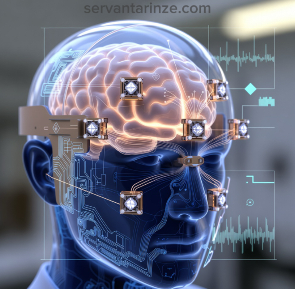
Speculation surrounding neural interfaces often outruns reality, and maintaining proportion is essential. Direct human brain mapping through NV arrays remains distant due to penetration limits, placement logistics, and signal attenuation across tissue layers. Yet discussions emerging from neurotechnology conferences increasingly treat diamond sensing as a candidate component in hybrid interface stacks. Not as the sole measurement layer, but as a near-surface contributor working alongside optical or electrical instrumentation.
This perspective reflects a broader shift toward modular sensing architectures. Instead of seeking a universal measurement solution, researchers assemble complementary sensing channels tuned to specific spatial and temporal sensitivities. NV platforms slot naturally into this framework, offering nanoscale resolution and ambient operability where other sensors introduce environmental or thermal constraints. Their contribution lies in versatility and integration potential rather than unilateral supremacy.
Ethical considerations inevitably accompany discussion of neural observation technologies. Greater spatial resolution introduces questions regarding cognitive privacy and responsible data stewardship. Although current NV implementations remain far from invasive or comprehensive monitoring capability, ethical discourse develops alongside capability trajectories rather than trailing behind them. The presence of quantum sensing within neuroscience dialogue therefore expands both technical and philosophical boundaries.
Beyond the Brain: Biological and Hybrid Sensing
Limiting NV relevance solely to neural systems would obscure its broader biological resonance. Cellular-scale thermometry, radical detection, and molecular environment sensing have already demonstrated feasibility through similar spin-resonance interactions. Observing intracellular magnetic or thermal variation contributes insight into metabolic and chemical dynamics, extending quantum sensing influence across biomedical investigation.
Hybrid bio-quantum interfaces represent another frontier drawing interdisciplinary curiosity. Integrating diamond-based sensing platforms with photonic or microfluidic structures allows simultaneous optical stimulation and magnetic observation. These configurations encourage experimental designs where stimulation and measurement operate in tightly coordinated cycles, deepening feedback loops between intervention and observation.
From a broader scientific perspective, a notable feature of this technological trajectory is the reduction of separation between abstract physics and biological immediacy. The same spin transitions described through Hamiltonians and resonance spectra become tools for probing living systems. That continuity reinforces the idea that quantum fundamentals are not isolated theoretical constructs but active contributors to empirical investigation across disciplines.
Applications will inevitably evolve as instrumentation matures and interdisciplinary familiarity increases. Whether NV sensing ultimately occupies niche specialization or widespread integration remains secondary to the current reality: it already provides experimentally meaningful insight where previous methods encountered observational blind spots. That contribution alone secures its place within the expanding toolkit of modern neuroscience and biological physics.
Where the Limits Still Stand: Constraints, Engineering Friction, and Practical Boundaries
Any conversation about sensing capability becomes unbalanced if limitations are treated as footnotes rather than structural realities. The enthusiasm surrounding diamond-based magnetometry sometimes obscures the fact that the physics enabling sensitivity simultaneously imposes operational constraints. These are not temporary inconveniences awaiting software updates. They arise from geometry, signal strength, and the interaction between biological environments and measurement apparatus. Understanding them is essential to interpreting experimental results responsibly.
The most persistent constraint is depth accessibility. Magnetic fields decay rapidly with distance, and neural currents are not exceptionally strong emitters. NV sensors excel when positioned extremely close to their source — micrometer or sub-millimeter regimes — but signal integrity diminishes sharply as separation increases. This places natural limits on non-invasive observation through intact cranial structures. Current demonstrations therefore emphasize tissue slices, cultured systems, or shallow-access models rather than full cortical imaging. Even ambitious ambient magnetometry pilots operate within carefully managed proximity parameters.
Scaling sensitivity introduces another tradeoff that laboratories continuously negotiate. Single-center measurements deliver exceptional spatial resolution but collect limited signal intensity, demanding extended integration periods or averaging strategies. Ensemble approaches aggregate signal strength across many centers, improving detection reliability at the cost of spatial specificity. This tension between resolution and sensitivity is not easily resolved; it reflects measurement fundamentals rather than instrumentation maturity. Researchers frequently tailor experimental configuration based on which dimension — spatial precision or signal robustness — carries greater relevance for the question under investigation.
Environmental Interference and Calibration Demands
External magnetic noise complicates observation more than theoretical sensitivity curves imply. Urban electromagnetic environments contain fluctuations from infrastructure, electronics, and natural geomagnetic variation. Shielding mitigates some disturbances, yet portable or ambient deployment requires sophisticated noise rejection techniques and statistical filtering. Maintaining calibration stability across experimental sessions becomes a nontrivial exercise in environmental management. Signal interpretation must therefore account for background behavior that may mask or distort biological signatures.
Temperature stability and optical alignment introduce further operational demands. Laser-driven readout requires precision that cannot be assumed once systems leave controlled laboratory settings. Photonic drift, vibration, or material inconsistencies may introduce measurement variance. These engineering factors seldom appear in theoretical discussions yet occupy considerable experimental effort. Much of quantum sensing progress stems not from altering spin physics but from refining instrumentation resilience against mundane physical disturbance.
Biological Integration Challenges
Working near or within biological material adds layers of complexity beyond sensor calibration. Optical illumination must remain within safe exposure ranges. Surface placement strategies must respect tissue viability. Mechanical positioning must avoid altering physiological behavior. These constraints transform sensor deployment into a multidisciplinary exercise involving materials science, optics, and biological protocol design. Even inert diamond substrates interact with experimental environments through thermal and mechanical pathways that must be understood rather than assumed negligible.
Signal interpretation also demands caution. Neural magnetic signatures overlap and propagate, meaning measured distributions reflect composite activity rather than isolated neuron behavior. Separating contributing sources requires modeling assumptions that introduce uncertainty. Increasing resolution reduces ambiguity but never eliminates it entirely. Observational clarity remains probabilistic rather than absolute, reinforcing the experimental nature of interpretation.
Ethical and Computational Realities
As sensing resolution improves, questions regarding data stewardship and interpretation responsibility follow closely. Extracting neural patterns carries implications extending beyond physics and engineering. Ethical frameworks surrounding consent, application boundaries, and interpretation transparency increasingly accompany technical development discussions. Responsible advancement requires these conversations to remain integrated with capability evolution rather than appended afterward.
Computational processing represents another quiet limitation. ODMR spectral fitting, vector reconstruction, and noise filtering generate data streams demanding sophisticated analysis pipelines. Extracting meaningful interpretation from raw sensing output relies on statistical modeling and often machine-assisted processing. As array sizes increase, computational infrastructure becomes an integral component of sensing architecture rather than a downstream convenience.
Despite these challenges, limitations should not be mistaken for stagnation indicators. They function as boundary markers guiding iterative refinement. Quantum sensing historically progresses through incremental engineering insight rather than singular conceptual breakthroughs. Each constraint navigated reshapes application feasibility and expands operational reach. Appreciating these realities strengthens experimental credibility and prevents interpretive overreach.
Beyond the Laboratory Horizon: Where Diamond Quantum Sensors Are Headed
Longitudinal assessment of nitrogen-vacancy sensing development indicates what stands out is not a single disruptive leap but a steady tightening of capability margins. Improvements in coherence engineering, photon collection efficiency, microwave control, and array fabrication have accumulated into a sensing platform that now operates at the boundary between laboratory instrumentation and translational neuroscience. The direction of travel is clear: greater parallelism, deeper integration with computational inference, and gradual movement toward deployment environments outside specialized facilities.
One trajectory attracting sustained investment is the development of scalable sensing arrays. Rather than relying on isolated sensing points, laboratories are constructing patterned ensembles capable of parallel acquisition across extended spatial regions. Increasing sensor density allows distributed field reconstruction that resembles imaging rather than point measurement. Engineering challenges remain substantial — optical uniformity, cross-talk minimization, synchronization — yet incremental progress has demonstrated feasibility. Multi-pixel NV magnetometry already supports spatial mapping experiments that were previously impractical with serial acquisition techniques.
Hybridization with Optical and Computational Systems
Sensing capability alone does not determine interpretive value. Future platforms are increasingly defined by their integration with optical microscopy and adaptive computational modeling. Coupling NV measurements with fluorescence imaging, for instance, enables correlation between magnetic activity and structural cellular features. These hybrid observation modes encourage richer experimental narratives, allowing researchers to contextualize magnetic signatures within biological architecture rather than treating them as abstract data distributions.
Artificial intelligence and statistical inference systems are simultaneously becoming embedded components of measurement pipelines. Pattern extraction from noisy magnetometry output demands interpretive assistance beyond manual inspection. Machine learning approaches are now applied to denoising, signal separation, and predictive modeling of neural field dynamics. This shift alters the nature of experimentation: the sensor and algorithm form a cooperative analytical instrument rather than independent stages of workflow.
Prospects for Neurotechnology and Interface Design
The conversation surrounding future application inevitably intersects with neurotechnology development. High-resolution, room-temperature sensing presents intriguing possibilities for noninvasive neural interface systems and diagnostic mapping tools. While speculative projections often overreach, grounded expectations point toward incremental integration into experimental neuroscience infrastructure first — refining disease models, validating pharmacological response, and informing implant placement strategies. The influence on clinical practice will likely emerge through gradual accumulation of validated use cases rather than abrupt technological displacement.
Another frontier lies in wearable or portable magnetometry configurations. Research initiatives exploring ambient-field operation without extensive shielding indicate movement toward mobility. Achieving stable sensing outside controlled environments would transform deployment possibilities, enabling longitudinal observation rather than episodic measurement. This remains technically demanding, but early demonstrations suggest the barrier is logistical rather than conceptual.
Physics as an Enduring Anchor
Amid forward-looking speculation, it remains useful to remember that progress is ultimately governed by physical constants rather than ambition. Spin coherence, photon statistics, and magnetic attenuation define the operational landscape within which innovation unfolds. Advances therefore reflect increasingly refined engagement with those constants rather than escape from them. This continuity anchors future projection in realism and protects discourse from drifting into technological romanticism.
Diamond defects sensing neuronal magnetism would once have sounded implausible outside specialist communities. Today, the idea is experimental routine. That shift did not occur through conceptual reinvention of quantum mechanics but through sustained engineering dialogue with physical law. The same dialogue will shape the next decade, gradually extending the reach of quantum sensing into domains not yet practical but increasingly approachable.
Conclusion
Discussions of quantum sensing frequently begin with abstract physical descriptors — coherence times, spin manipulation, resonance shifts — but its relevance becomes unmistakable when those abstractions begin interacting with biological systems. Nitrogen-vacancy centers represent one of the clearest demonstrations that quantum physics is not confined to vacuum chambers or dilution refrigerators. A defect in a diamond lattice, engineered and interrogated correctly, can register electromagnetic signatures generated by living neural tissue. That transition from principle to interaction is where the field gains its real weight.
The physical mechanisms enabling this interaction are neither mysterious nor speculative. Spin-dependent fluorescence, microwave-driven transitions, and Zeeman splitting are well-established elements of quantum control. Their significance lies in their resilience at ambient conditions and nanoscale locality. When combined with careful optical and microwave engineering, these properties create a sensing platform that is both delicate in sensitivity and robust in operation. It is this combination that makes NV magnetometry uniquely positioned between laboratory physics and applied neuroscience.
Observing the field’s trajectory, one sees a pattern familiar across technological history. Early demonstrations emphasize feasibility. Subsequent iterations refine measurement stability and interpretive confidence. Only later do integration and accessibility reshape surrounding disciplines. NV sensing currently resides within that refinement phase — strong enough to reshape experimental practice, not yet diffuse enough to redefine clinical infrastructure. Recognizing that stage prevents exaggerated expectation while still appreciating the scale of potential transformation underway.
A broader conceptual implication also emerges. Mapping neural magnetic fields using quantum defects reframes how knowledge of the brain is pursued. Instead of inserting probes or relying on indirect metabolic indicators, measurement approaches are shifting toward minimally intrusive observation of intrinsic electromagnetic behavior. The implications extend beyond instrumentation — they influence how neuroscientists conceptualize observation itself, altering the balance between intervention and perception.
From a broader vantage point, nitrogen-vacancy sensing illustrates the evolving relationship between quantum engineering and everyday scientific inquiry. Technologies once categorized as frontier physics now function as investigative tools across disciplines. Chemistry, biology, and materials science increasingly rely on quantum-enabled measurement without necessarily foregrounding the quantum origin of the capability. This quiet integration may ultimately represent the field’s most enduring impact.
What remains ahead is incremental rather than theatrical. Enhanced photon collection, longer coherence preservation, denser sensing arrays, and improved computational interpretation will continue to push sensitivity and scale. Each adjustment slightly expands what can be observed and slightly reduces ambiguity in interpretation. Over time, these cumulative adjustments reshape possibility. The narrative of progress in quantum sensing is written not through singular revolutions but through steady recalibration of experimental reach.
Nitrogen-vacancy centers do not map thoughts, and they do not replace established neuroimaging modalities. What they provide is an additional observational channel — one rooted in quantum physics — capable of revealing magnetic dynamics previously difficult to resolve. That contribution is substantial enough to influence experimental methodology and subtle enough to integrate alongside existing tools rather than displace them.
The broader lesson extends beyond this specific technology. When fundamental physics becomes instrumentation, the boundary between theoretical understanding and empirical exploration dissolves. NV centers exemplify that dissolution. They transform lattice imperfections into sensors, and in doing so they demonstrate how quantum mechanics increasingly participates in interpreting the living world rather than merely describing the microscopic one.
This intersection of condensed matter physics, optical engineering, and neuroscience is unlikely to remain niche. As fabrication matures and interpretive frameworks strengthen, diamond-based quantum sensing will continue migrating toward wider adoption. Whether through refined experimental mapping, biomedical diagnostics, or hybrid neurotechnology systems, its influence will be measured not in spectacle but in accumulated insight.
That trajectory reinforces a quiet but powerful truth: advances in physics rarely end within physics. They propagate outward, altering how other domains observe themselves. Nitrogen-vacancy centers are one instance of that propagation, translating quantum spin behavior into biological understanding — a reminder that the most consequential technologies often emerge from the most unassuming physical irregularities.
References
- Doherty, Marcus W., Neil B. Manson, Paul Delaney, Fedor Jelezko, Jörg Wrachtrup, and Lloyd C. L. Hollenberg. 2013.
“Nitrogen-Vacancy Colour Centres in Diamond.”
Nature Physics 9: 139–143.
View Source
↩ - Rondin, L., J.-P. Tetienne, T. Hingant, J.-F. Roch, P. Maletinsky, and V. Jacques. 2014.
“Magnetometry with Nitrogen-Vacancy Defects in Diamond.”
Reports on Progress in Physics 77 (5).
View Source
↩ - Taylor, J. M., P. Cappellaro, L. Childress, L. Jiang, D. Budker, P. R. Hemmer, A. Yacoby, et al. 2008.
“High-Sensitivity Diamond Magnetometer with Nanoscale Resolution.”
Nature Physics 4: 810–816.
View Source
↩ - Hämäläinen, M., R. Hari, R. J. Ilmoniemi, J. Knuutila, and O. V. Lounasmaa. 1993.
“Magnetoencephalography — Theory, Instrumentation, and Applications.”
Reviews of Modern Physics 65 (2): 413–497.
View Source
↩
Frequently Asked Questions About NV-Center Quantum Sensors & Brain Mapping (FAQ)
What is a nitrogen-vacancy center in a diamond?
A nitrogen-vacancy (NV) center is a tiny defect in a diamond lattice where a nitrogen atom replaces a carbon atom next to an empty site. This defect hosts an electron spin that can be controlled and read optically, allowing it to act as a highly sensitive quantum sensor for magnetic fields, temperature, and electric fields at room temperature.
How can NV centers detect activity in the human brain?
Neurons generate tiny magnetic fields when electrical currents flow during signaling. NV sensors detect these fields through shifts in their spin resonance frequencies. By measuring fluorescence changes under optical excitation, researchers can map neural magnetic activity with high spatial and temporal precision.
Are diamond quantum sensors better than EEG or fMRI?
They are not replacements but complementary tools. EEG offers excellent timing resolution but coarse spatial detail, while fMRI provides detailed spatial mapping but slow temporal response. NV magnetometry promises finer localization than EEG and faster dynamics than fMRI, though it remains largely experimental for in-vivo human use.
Do NV quantum sensors require cryogenic cooling?
No. One of their major advantages is stable quantum coherence at room temperature. Unlike SQUID magnetometers that require liquid helium cooling, NV sensors operate under ambient conditions, enabling portable and biologically compatible experimental setups.
What limits NV-based brain mapping today?
Current constraints include sensing depth, signal strength from single neurons, environmental magnetic noise, and integration challenges for non-invasive deployment. Most demonstrations remain in tissue samples or shallow biological environments, with ongoing work addressing scalability and sensitivity.